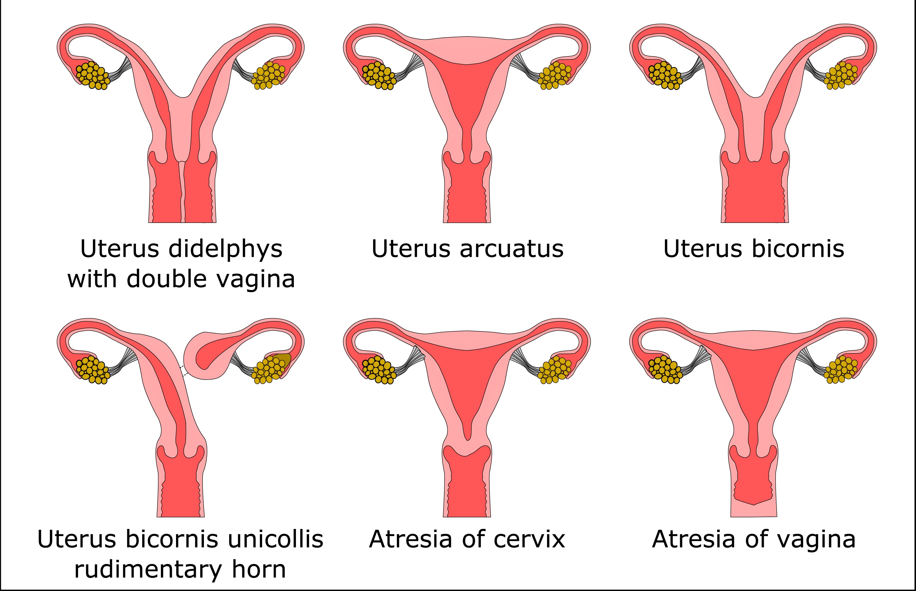
Uterine Prolapse Surgery in a woman with Uterine Didelphy
A 60-year-old woman presented to Dr. Miklos & Moore a complaint of uterus prolapse and uterus didelphy (i.e. two uteruses). She was diagnosed with a didelphic uterus approximately 30 years ago. She was seen by 3 gynecologists, in Atlanta, all of which either refused to do her prolapse surgery or recommended a hysterectomy. She informed the surgeons she would not settle for a hysterectomy, as she wanted to preserve her uterus. She was finally referred to the Mayo Clinic and Emory University.
She then decided to see Dr. Miklos for symptoms of prolapse, pressure, as well as stress urinary incontinence. Dr. Miklos explained uterine preservation was indeed an option and this was not the first time that he had performed a hysteropexy in a woman with uterus didelphy.
On examination, she was noted to have two vaginal canals (Figure 1), two cervixes (Figure 2), and two uteruses. She was also diagnosed with pelvic floor relaxation in the form of a cystocele, rectocele, and uterine prolapse x 2. Testing revealed stress urinary incontinence. A thorough discussion as to the risks and benefits of surgery were discussed. Most importantly, Dr. Miklos informed the patient that he could and would meet her independent decision to save her uterus and would not perform a hysterectomy.
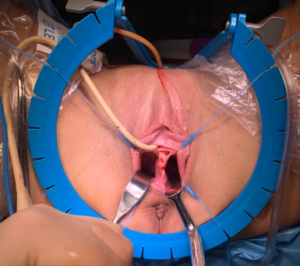
Figure 1: Two Vaginal Canals
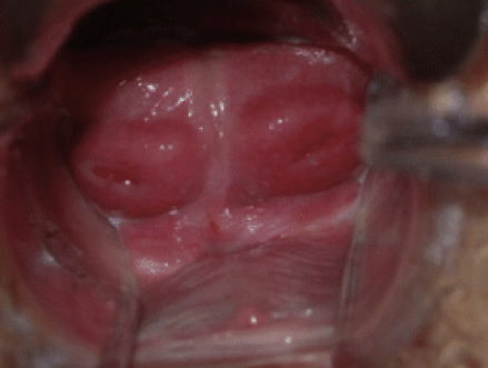
Figure 2: Two Cervixes
The patient had her surgery including: 1) laparoscopic hysteropexy (aka sacrocolpohysteropexy) 2) paravaginal repair, 3) Burch urethropexy (for cough urine leakage), & 4) a rectocele repair. He also performed (per her request) a vaginal rejuvenation or “tightening surgery.” The surgery was performed without complications with an estimated blood loss of less than 25 mL. There were no intraoperative complications or immediate postoperative complications and she was discharged less than 48 hours after the surgery. A photo of the patient’s two uteruses can be seen in Figure 3. Dr. Miklos performs the hysteropexy procedure in Figure 4.
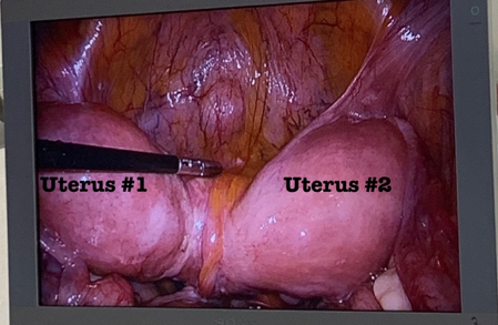
Figure 3: Laparoscopic view of Uterus Didelphy (Two Uteruses)
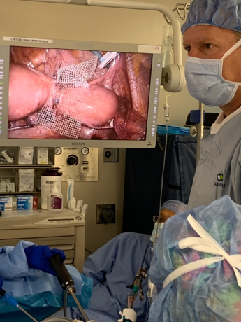
Figure 4: Dr. Miklos performs the hysteropexy